The Evolution of Surgical Training: Beyond the Cadaver with 3D Printing
Introduction: A New Standard in Surgical Proficiency
Traditional surgical training has long relied on a combination of 2D imaging, generic plastic models, and cadaveric specimens. However, these methods face increasing challenges: cadavers are expensive and lack specific pathologies, while 2D scans require a “mental leap” to 3D reality.
In 2025, 3D printing has transitioned from an experimental “nice-to-have” to a clinical standard. By converting DICOM data from CT/MRI scans into high-fidelity physical models, surgeons can now rehearse complex procedures on a patient’s exact anatomy before entering the OR.
1. From Visual to Haptic: Material Breakthroughs
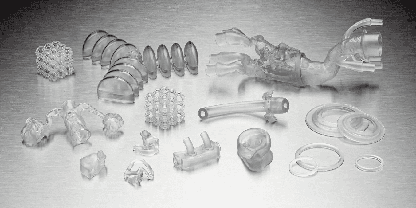
The most significant update since the original article is the development of biocompatible and “tissue-mimicking” resins.
- Elastic and Flexible Resins: New materials like Formlabs BioMed Elastic 50A and BioMed Flex 80A allow for the creation of soft tissue models that respond to sutures, incisions, and catheter navigation like real human tissue.
- Multi-Material Printing: Advanced systems can now print “hybrid” models—combining rigid bone-like structures with soft vascular networks in a single print—providing the realistic resistance (haptic feedback) necessary for training.
2. Case Studies: Precision Cardiology (Formlabs Insights)
Recent medical literature highlights how 3D printing is specifically solving “blind-spot” challenges in cardiology:
- Left Atrial Appendage Occlusion (LAAO): A 2024 study showed that 3D-printed models were more accurate for sizing occlusion devices than standard 2D/3D CT scans. Using these models significantly reduced “LAA leaks,” a common post-operative complication.
- Congenital Heart Disease (CHD): For pediatric surgeons, 3D models of tiny, malformed hearts provide a spatial understanding that even Virtual Reality (VR) struggles to replicate. 76% of practitioners in a recent study reported that physical 3D models directly increased their confidence before CHD surgeries.
- Hybrid Procedures: Surgeons are now using 3D models to simulate “stay-suture” techniques in real-time, acting as a “fail-safe” for complex device deployments in small patients.
3. Measurable Clinical & Economic Outcomes
The shift to 3D printing isn’t just about better training; it’s about better hospital metrics. Recent data (2024-2025) confirms:
- Reduced OR Time: Pre-surgical rehearsal on 3D models can reduce operating room time by up to 22%. In a hospital setting, saving 30 minutes of OR time can equate to thousands of dollars in cost savings.
- Lower Complication Rates: Surgeons who practice on patient-specific models report lower intraoperative blood loss and higher procedural accuracy, particularly in maxillofacial and orthopedic surgeries.
- Cost Efficiency: While a cadaveric lab can cost upwards of $15,000 per session, in-house FDM or SLA 3D printing can produce high-quality training phantoms for under $400, making repetitive practice economically viable.
4. Patient Communication and Informed Consent
A hidden benefit of the 3D printing revolution is the “Patient-Centered” approach. 3D models serve as a powerful tool for:
- Informed Consent: Patients and families report a significantly higher understanding of their condition when holding a 3D model compared to looking at a screen.
- Anxiety Reduction: Studies show that when patients visualize exactly how a “patch” or “stent” will be placed in their own heart or bone, pre-surgical anxiety is markedly reduced.
Conclusion: The Future of the “Point-of-Care” Lab
The outdated model of ordering anatomical models from external vendors is being replaced by Point-of-Care (POC) 3D Printing. With the arrival of faster, more accessible printers like the Form 4B, hospitals are now running 24/7 labs within their own radiology departments—turning a morning CT scan into a lunchtime surgical rehearsal model.
Key Sources:
Formlabs Medical Literature Roundup (Cardiology)
2026 Systematic Review (EMJ Radiology)
MDPI Review (2024)